3 tips for a healthy feline diet
9 Aug 2022
Vet Image Solutions, passionate about ultrasound.
Below is a feline uterus, removed during a spay. The ovaries can be seen at the ends of the two horns.
AN EXAMPLE OF ACOUSTIC SHADOWING
One of the most common questions is “how can I optimise my image on the [insert your ultrasound machine name here]?” It’s a great question, but it’s also one of the most difficult to answer quickly.
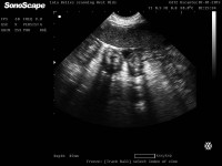
Sian Davis of Lola Belles Mobile Pet Scanning in the West Midlands recently shared a scan of a closed pyo on the Facebook group. She had been called out by a breeder who had been sent home by the local vet, but she was sure that something wasn't right with her bitch.

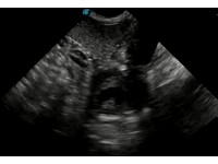
The Vet Image Solutions Facebook group is a fantastic learning resource for those looking to learn more about canine reproduction and scanning. Recently, Jennie from Lincoln Canine Scanning shared this scan with the following comment:
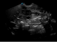
English Bulldog 43 days?? Water pup here, could see it better on the screen but moved just as took the picture - abdominal wall seen with fluid around anterior abdominal wall.
This quick video clip illustrates the difference between the appearance of gestation sacs and intestines on a canine or feline ultrasound scan. Note the 'hamburger' shape of the intestines, which would appear tubular with rotation of the probe by 90 degrees. The pregnancy sacs, on the other hand, are circular at this early stage (30 days) and would maintain their distinct borders even with rotation of the transducer.